Hysterosalpingography
Hysterosalpingography (HSG), also known as uterosalpingography,[1] is a radiologic procedure to investigate the shape of the uterine cavity and the shape and patency of the fallopian tubes. It is a special x-ray using dye to look at the womb (uterus) and Fallopian tubes[2] It injects a radio-opaque material into the cervical canal and usually fluoroscopy with image intensification. A normal result shows the filling of the uterine cavity and the bilateral filling of the fallopian tube with the injection material. To demonstrate tubal rupture, spillage of the material into the peritoneal cavity needs to be observed. It has vital role in treatment of infertility especially in case of fallopian tube blockage.
| Hysterosalpingography | |
|---|---|
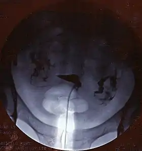 A normal hysterosalpingogram. Note the catheter entering at the bottom of the screen, and the contrast medium filling the uterine cavity (small triangle in the center). | |
| Other names | Uterosalpingography |
| ICD-9-CM | 87.8 |
| MeSH | D007047 |
| MedlinePlus | 003404 |
.jpg.webp)
Uses
HSG is considered a diagnostic procedure. It is used in workup of infertile women to access the patency of fallopian tubes, access the competency of cervix or congenital abnormality of the uterus in multiple miscarriages, to access patency of fallopian tubes after surgery or tubal ligation or before reversal of tubal ligation. Rarely, HSG is used to access the integrity of caesarean scar.[3]
HSG may also have therapeutic benefits for infertility treatment. When oil-based contrast is used rates of pregnancy increase by about 10% compared to water-based contrast.[4] A meta-analysis revealed 3.6 times greater odds (OR = 3.6) of pregnancy with oil-based contrast compared to no hysterosalpingography.[5] This effect is thought to be due to tubal flushing with the oil-based contrast rather than the imaging procedure itself.
HSG is contraindicated during menstruation, pregnancy, or any unprotected sexual intercourse during the menstrual cycle, any purulent discharge from the vagina, or was diagnosed as pelvic inflammatory disease at previous six months. Those with hypersensitivity to contrast is relatively contraindicated in HSG.
Procedure
Either high osmolar contrast material (HOCM) or low osmolar contrast material (LOCM) can be used. 10 to 20 ml of LOCM can be used at a concentration of 270 to 300 mg/ml. The contrast media should be prewarmed to room temperature before administered into the cervix so as to prevent spasm of fallopian tubes. 5Fr to 7Fr hysterosalpingogram balloon catheter can be used. Margolin hysterosalpingraphy cannula is used if the cervix is narrow or stenosed.[3] HSG appointment is usually made during the 4th to 10th days of regular mentrual cycle. The subject should not undergone any sexual intercourse before HSG. Anxious subjects may need painkillers or other medications. Informed consent should be taken before the procedure.[3]
The subject lie down on table in supine position with legs flexed and abducted. Vulva is cleaned with chlorhexidine or normal saline. A speculum is inserted to the vagina with the help of sterile jelly, and the cervix is exposed. The cervical opening is identified using a bright light. The HSG catheter is then inserted into the cervical canal. Occasionally, Vulsellum forceps may be used to hold the cervical lips open. If cervical weakness is suspected, the catheter should be left inside the lower cervical canal.[3] Air bubbles should be expelled from the syringe and the catheter, otherwise it will cause confusion of interpretations on HSG. Contrast medium is injected slowly into the uterine cavity with intermittent fluoroscopic screening. If there is no spills from bilateral fallopian tubes bilaterally, intravenous buscopan and glucagon can be given to relieve spasm of fallopian tubes.[3] Opiates should not be given as it may increase pain because of increased smooth muscle contractions.[3]
Images are taken to demonstrate the filling of endometrial cavity, full view of the fallopian tubes demonstrating spillage contrast into peritoneum or the extent of block if there is no spilling, and a delayed view if there is abnormal cavities (lovule) within. Subject may have vaginal spotting for one to two days with pain and may persists for up to two weeks. Some centres routinely give prophylactic antibiotics before subject is allowed home.[3]
The procedure involves X-rays. It should be done in the follicular phase of the cycle.[6] Using catheters, an interventional radiologist or specifically trained radiographer can open tubes that are proximally occluded.
The test is usually done with radiographic contrast medium (dye) injected into the uterine cavity through the vagina and cervix. If the fallopian tubes are open the contrast medium will fill the tubes and spill out into the abdominal cavity. It can be determined whether the fallopian tubes are open or blocked and whether the blockage is located at the junction of the tube and the uterus (proximal) or whether it is at the end of the fallopian tube (distal).
The HSG can be painful, so analgesics may be administered before and/or after the procedure to reduce pain. Many doctors will also prescribe an antibiotic prior to the procedure to reduce the risk of an infection.[3]
Complications
Complications of the procedure include infection,[2] allergic reactions to the materials used,[2] intravasation of the contrast material, pain during the procedure, nausea, vomiting, and headache. Some subjects may develop neurogenic shock during the inflation of balloon in the cervical canal.[3]
History
For the first HSG, Carey used collergol in 1914. Lipiodol was introduced by Sicard and Forestier in 1924 and remained a popular contrast medium for many decades.[7] Later, water-soluble contrast material was generally preferred as it avoided the possible complication of oil embolism.
Follow up
If the HSG indicates further investigations are warranted, a laparoscopy, assisted by hysteroscopy, may be advised to visualise the area in three dimensions, with the potential to resolve minor issues within the same procedure.
See also
References
- "Hysterosalpingography (Uterosalpingography)". RadiologyInfo. June 8, 2016.
- "Hysterosalpingography: MedlinePlus Medical Encyclopedia". medlineplus.gov. Retrieved 2019-05-06.
- Watson N, Jones H (2018). Chapman and Nakielny's Guide to Radiological Procedures. Elsevier. pp. 163–166. ISBN 9780702071669.
- Dreyer, Kim; Rijswijk, Joukje van; Mijatovic, Velja; Goddijn, Mariëtte; Verhoeve, Harold R.; Rooij, Ilse A.J. van; Hoek, Annemieke; Bourdrez, Petra; Nap, Annemiek W. (2017-05-18). "Oil-Based or Water-Based Contrast for Hysterosalpingography in Infertile Women". New England Journal of Medicine. 376 (21): 2043–2052. doi:10.1056/nejmoa1612337. PMID 28520519.
- Wang, Rui; Watson, Andrew; Johnson, Neil; Cheung, Karen; Fitzgerald, Cheryl; Mol, Ben Willem J.; Mohiyiddeen, Lamiya (October 15, 2020). "Tubal flushing for subfertility". The Cochrane Database of Systematic Reviews. 10: CD003718. doi:10.1002/14651858.CD003718.pub5. ISSN 1469-493X. PMID 33053612. S2CID 222421134.
- Baramki T (2005). "Hysterosalpingography". Fertil Steril. 83 (6): 1595–606. doi:10.1016/j.fertnstert.2004.12.050. PMID 15950625.
- Bendick A. J. (1947). "Present Status of Hysterosalpingography". Journal of the Mount Sinai Hospital, New York. 14 (3): 739–742. PMID 20265114.
External links
- HSG Test - HSG Test : Cost, Results, Expert Advice, Preparation
- HSG - HSG Test -HSG Film Advice, Preparation
| X-ray/ Radiography | |||||||||||||
|---|---|---|---|---|---|---|---|---|---|---|---|---|---|
| MRI | |||||||||||||
| Ultrasound | |||||||||||||
| Radionuclide |
| ||||||||||||
| Optical/Laser | |||||||||||||
| Thermography |
| ||||||||||||
| Target conditions | |||||||||||||
| |||||||||||||